For a long time if you had significant back pain then imaging was considered the first step towards finding the right care and a solution. Science is very clear now that this is not the case. That, in fact, imaging can be detrimental to the cause.
Research shows that when people experience back pain the population at large believes that to get the best outcome from the health system they need two things: imaging of their lower back (1) and a name for their painful tissue ~ a diagnosis (2).
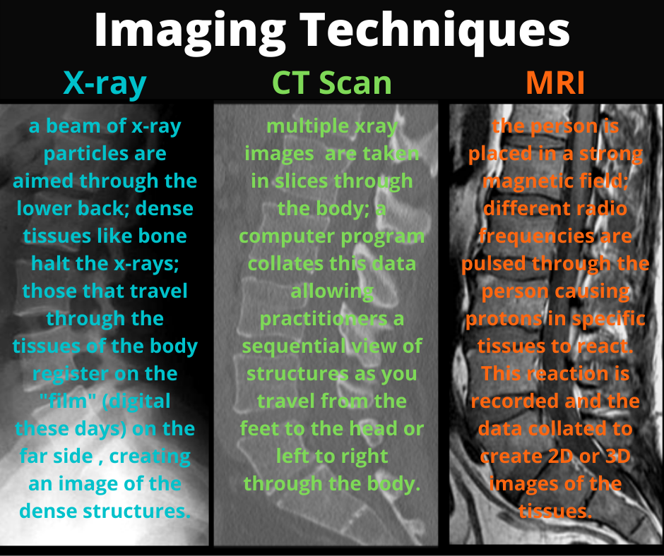
Patient beliefs about back imaging (1):
They believe that having this imaging will mean that appropriate treatment can be given and the problem can be cured. I understand that. I often tell people to collate all the data so they can then make good choices about their health, but does imaging provide us with useful data about back pain?
We know that only 1% of new onset low back pain cases that present to a primary care practitioner (i.e. a GP, chiro or physio; not to a hospital) have a specific cause (3). It is really rare to have a nasty reason for your back pain, but your primary care practitioner is trained to detect these causes and refer you on (possibly for imaging) where necessary. The vast majority of people experiencing lower back pain fall into the category known as “Non-specific Low Back Pain” (a.k.a LBP with no indication of underlying pathology). Imaging is most useful for detecting pathological changes, but if only 1% of low back pain cases presenting to primary care practitioners have pathological reasons for their pain, then we can see why routine imaging for low back pain is not recommended.
There is a link between the bony changes and disc changes we see on imaging and low back pain. However, if you were to do imaging on a randomly selected citizen of a similar age who is pain free, you are likely to see bony and soft tissue changes that are much the same as the symptomatic person because these changes are normal and increase in incidence with time in all of us (4). To quote David Butler, they are only “the kisses of time”.


Unfortunately, the consequence of having imaging done for people experiencing low back pain is that when practitioners receive an imaging report from a radiologist that has identified such changes in the spine in a similar region as the person’s pain, then the practitioner is likely to link the two findings and tell the patient that the changes found on imaging are the source of the person’s pain.
We now know that identifying these changes with imaging and naming them for the person actually does them a disservice because:
[Remember pain is a tool the brain uses to keep us safe, not an indicator of tissue damage. To read more on this please click on this link to read our blog on PAIN]
(a) a worse prognosis because of what the patient now believes about their back pain (which is that they’re now set to have back problems for life) (5),
(b) and greater levels of intervention (opioids/lyrica (pregabalin), injections, surgeries) (2,6).
To quote Jan Hartvigsen in the Lancet series on Low Back Pain ~
In fact Lemmers et. al. published a review of the literature in 2019 (6) which established that, from the perspective of health care systems and governments,
It’s no wonder the Australian Government and governments around the world are producing guidelines (for their health care providers and their people) advising against routine imaging for non-specific low back pain (8,9).
Instead, the Australian Government Department of Health advises, the best care and best outcomes for people experiencing non-specific low back pain are achieved by receiving conservative spine management (including spinal manipulation) which you’ll get from your chiropractor or physiotherapist.